WGN Medical Watch – 1/30/2013 – Forecasting brain tumors

Forecasting brain tumors

Predicting brain tumors like weather forecasters predict an upcoming storm. For patients, that means they can better prepare and make choices for how to better protect themselves.

WGN meteorologist Tom Skilling surrounds himself with mathematical data. Wind speed, barometric pressure, temperature rise and fall. Armed with that knowledge, he creates the daily and weekly forecast.

In this lab, researchers are doing the exact same thing, except they are forecasting how quickly a brain tumor will erupt and spread. It lets patients know if they should ditch the raincoat for a stronger protector from the elements.

Prof. Kristin Swanson, Northwestern Medicine Brain Researcher: “So if a patient comes in and gets some new treatment, you say ‘I predicted the disease was going to be this big at some future time point,’ and the disease was only this big, then that difference is a measure of how well the treatment worked. And that difference is actually very predictive of how well a patient does in overall survival.”

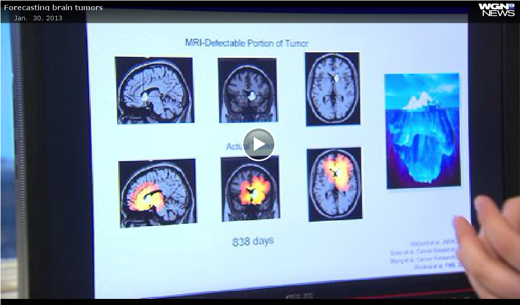
Look at this image. Think of the white spot as an iceberg, the part you can see above water. Then look at the image below. The red represents the true size — the growth and tumor spread through the brain, like the iceberg below the water.

Prof. Kristin Swanson: “It’s all about seeing what you can’t see. The imaging doesn’t tell you the whole answer so how can you infer where the disease is?”

MRI is good but limited. Instead, researchers at Northwestern Medicine are using a mathematical model comparing the rate of growth on MRI and analyzing cell density.

Prof. Kristin Swanson: “If you’ve got two pre-treatment MRI’s, you can tune the mathematical model to each patient. One of the nice things about this tool set is that you can not only predict what the disease extent is, but you can say, ‘Hey, this is what this patient looks like today, this is what I think the patient’s going to look like in six weeks, this is what I think the patient’s disease is going to look like in six months.’ And when you combine all that information together, you can generate a baseline against which to compare treatments.”

Plotting points on a graph, in this case the line represents how large and fast the tumor will grow. But then treatment is started. Now look at the red box below the line. It represents how much smaller the mass was from the original prediction. The cancer is no longer rising and growing, but the angle of progression is shrinking … the therapy is working. It tells doctors to stay the course.

Prof. Kristin Swanson: “They need to know whether the treatment that they are on is doing the right thing. So that’s one of the nice things about this tool is that it says, ‘Hey, these patients are actually getting a lot of bang for their buck, they’re getting a ton from this given therapy, where these patients over here, they may not be getting as much.’”

The ultimate goal is to accurately predict exactly when to change therapies. Currently, some patients stick with what they know without opting for a clinical trial when it may save their lives, others give up on the strongest standard therapy too soon. The researchers are hoping prediction will mean life extension.

https://wgntv.com/2013/01/30/forecasting-brain-tumors/

Donate to the
Swanson Lab


Forecasting Brain Tumors Like a Storm

FORECASTING BRAIN TUMORS LIKE A STORM

New method is first to predict brain cancer outcome and quickly show if therapy is effective

CHICAGO — The critical question shortly after a brain cancer patient starts treatment: how well is it working? But there hasn’t been a good way to gauge that.

Now Northwestern Medicine researchers have developed a new method — similar to forecasting storms with computer models — to predict an individual patient’s brain tumor growth. This growth forecast will enable physicians to rapidly identify how well the tumor is responding to a particular therapy. The approach allows a quick pivot to a new therapy in a critical time window if the current one isn’t effective.

The study is based on 33 patients with glioblastoma, the most common and aggressive form of brain cancer. The paper will be published Jan. 23 in the journal PLOS ONE.

“When a hurricane is approaching, weather models tell us where it’s going,” said senior author Kristin Swanson, professor and vice chair of research for neurological surgery at Northwestern University Feinberg School of Medicine. “Our brain tumor model does the same thing. We know how much and where the tumor will grow. Then we can know how much the treatment deflected that growth and directly relate that to impact on patient survival.”

Swanson also is a member of the Northwestern Brain Tumor Institute and the Robert H. Lurie Comprehensive Cancer Center of Northwestern University. Maxwell Neal, lead author, is a post-doctoral researcher in bioengineering at the University of Washington.

The method will advance brain tumor treatment, Swanson said, by helping distinguish effective treatments from ineffective ones and enabling clinicians to optimize treatment plans on a patient-by-patient basis.

Muddy Zone Right After Treatment

“There is this muddy zone right after the first round of treatments when it’s hard for the clinician to know whether to change therapy because she doesn’t have the metrics that correlate to outcome,” Swanson said. “The doctor can’t yet gauge how much it helped.”

If the doctor determines the treatment isn’t effective, she can try a different type of treatment or help the patient enroll in a clinical trial with a new drug being tested. The information also is helpful to the patient.

“The patient wants to know the therapy is doing something for them,” Swanson said. “On the flip side, if the therapy isn’t helping, then it may not be worth the side affects he is enduring.”

Not All Brain Tumors are the Same

Brain cancer patients are in great need of an approach to find optimal personalized treatments.

Brain tumors vary in their growth rate, shape and density but existing methods for measuring a treatment’s impact ignore this variation. The methods (and thus physicians) cannot distinguish between a patient with a fast-growing tumor that responds well to treatment and a patient with a slow-growing tumor that responds poorly.

By using a personalized, patient-specific approach that accounts for tumor features such as 3-dimensional shape, density and growth rate, the new Northwestern method can make this distinction.

Is it Working? How the Model Forecasts Growth and Measures Effectiveness

To measure a treatment’s effectiveness, the scientists performing the study created a unique computer model of each patient’s tumor and predicted how it would grow in the absence of treatment, explained Neal.

The prediction model was based on the MRI scans that the patient received on the day of diagnosis and on the day of surgery. The difference between these two scans enabled researchers to estimate how fast the tumor was growing along with the density of tumor cells throughout the brain.

Researchers then scored the effectiveness of the patient’s treatment by comparing the size of the patient’s tumor after treatment to the model-predicted size if untreated.

“The study demonstrated that higher-scoring patients survived significantly longer than lower-scoring patients and their tumors took significantly longer to recur,” Neal said. “The score can guide clinicians in determining the effectiveness of the therapy.”

Northwestern researchers hope to make the computer model an iPad app or offer it on a website where a clinician can simply enter a patients’ MRI data to calculate the response score.

The research was supported by the National Cancer Institute of the National Institutes of Health, grants R01 CA164371, R01 NS 060752, U54 CA143970. In addition, the research was funded by the McDonnell Foundation, the Brain Tumor Funders Collaborative, the University of Washington Academic Pathology Fund, and the James D. Murray Endowed Chair.

NORTHWESTERN NEWS: https://www.eurekalert.org/pub_releases/2013-01/nu-fbt011813.php

Kristin R. Swanson named Professor and Vice Chair of Research for Neurological Surgery

After 12 years at the University of Washington, Kristin Rae Swanson, PhD, has been named professor and vice chair of research for neurological surgery at Northwestern University Feinberg School of Medicine.

As part of a recruitment effort to expand the Northwestern Brain Tumor Institute (NBTI), Kristin Rae Swanson, PhD, has been named professor and vice chair of research for the Department of Neurological Surgery effective October 22, 2012.

Swanson comes to Feinberg from the University of Washington, where she served as the James D. Murray Endowed Chair of Applied Mathematics in Neuropathology as part of the Nancy and Buster Alvord Brain Tumor Center. During her 12-year career there, she led a well-funded research effort pioneering the field of mathematical neuro-oncology as a novel means to generate personalized medicine approaches for primary brain tumors. Swanson has a talent for developing collaborative networks comprised of strong multidisciplinary researchers, scientists, clinicians, and trainees, which will strengthen the Brain Tumor Institute’s research endeavors.

“I am thrilled to be joining the Northwestern Brain Tumor Institute at such an exciting time of growth and opportunity,” Swanson said. “The institutional and community investment in growing the NBTI is astounding and I am delighted to be part of this exceptional group. I know my lab will contribute to this growth through the integration of our science into the clinical and research fabric of the Northwestern community.”

As vice chair for research, Swanson’s mentoring skills will be invaluable. In 2010, she was honored with the University of Washington Research Mentor of the Year Award. Swanson is also a member of multiple national organizations, including the American Association for Cancer Research, the Society for Mathematical Biology, the Society for Neuro-Oncology, and the Society for Nuclear Medicine.

“Dr. Swanson has distinguished herself as a leading authority in the area of mathematical models of gliomas and her research efforts will be a tremendous asset to the Northwestern scientific community,” said James Chandler, MD, surgical director of the NBTI.

See the announcement here

Presentations at 2012 Annual SNO Meeting

Ten abstracts including 9 posters and 1 talk from the Swanson research lab have been accepted for presentation at the annual Soceity for NeuroOncology meeting in Washington D.C. Wide-ranging and novel results include clinically translational applications of patient-specific mathematical modeling using IDH-1, stem cell transplant therapy, radiation therapy optimization and predictive outcomes based on extent of surgery.

See selected abstracts in the special issue of Neuro-Oncology here

https://neuro-oncology.oxfordjournals.org/content/14/suppl_6.toc

Discriminating time to progression and survival using a response metric tuned to patient-specific glioblastoma kinetics

New manuscript published in PLoS ONE (in press).

Discriminating time to progression and survival using a response metric tuned to patient-specific glioblastoma kinetics

Neal ML, Trister AD, Cloke T, Sodt R, Ahn S, Baldock AL, Bridge CA, Boone A, Rockne R, Swanson KR.

Accurate clinical assessment of a patient’s response to treatment for glioblastoma multiforme (GBM), the most malignant type of primary brain tumor, is undermined by the wide patient-to-patient variability in GBM dynamics and responsiveness to therapy. Using computational models that account for the unique geometry and kinetics of individual patients’ tumors, we developed a method for assessing response that discriminates progression-free and overall survival following therapy for GBM. Applying these models as untreated virtual controls, we generate a patient-specific “Days Gained” response metric that estimates the number of days a therapy delayed imageable tumor progression. We assessed treatment response in terms of Days Gained scores for 33 patients at the time of their first MRI scan following first-line radiation therapy. Based on Kaplan-Meier analyses, patients with significant treatment response (characterized by Days Gained scores of 100 or more) had improved progression-free survival and improved overall survival. Our results demonstrate that the Days Gained response metric calculated at the routinely acquired first post-radiation treatment time point provides prognostic information regarding progression and survival outcomes. Applied prospectively, our model-based approach has the potential to improve GBM treatment by accounting for patient-to-patient heterogeneity in GBM dynamics and responses to therapy.

Quantifying the role of angiogenesis in malignant progression of gliomas

New manuscript published in Cancer Research E-pub ahead of print

Cancer Research 2011 Sep 7.

Quantifying the role of angiogenesis in malignant progression of gliomas:

In silico modeling integrates imaging and histology.

Swanson KR, Rockne RC, Claridge J, Chaplain MA, Alvord EC Jr, Anderson AR.

Although commonly attributed to molecular-genetic factors such as the accumulation of genetic mutation, this study combines mathematical modeling with experimental and clinical data for human gliomas to suggest that changes in cell kinetics are not necessary to generate the imaging and histological features of malignant progression seen in vivo. Model predictions are validated against imaging and histological data for 3 GBM patients.

NIH R01 “UVIC” Grant Award

Untreated Virtual Imaging Control (UVIC)

funded by the National Institutes of Health (www.nih.gov).

The UVIC grant proposal integrates mathematical modeling of tumor proliferation and invasion with advanced cancer imaging methods. The goals of our project are twofold:

1) To impact current clinical challenges with treatment of gliomas

2) provide tools for the development of new therapies for these challenging cancers.

Our first goal is to develop image-based response metrics based on the growth kinetics of each patient’s tumor, as seen on both anatomical imaging (MR) and functional imaging (PET and advanced MR). We will use mathematical modeling to develop a patient-specific UVIC that quantifies the dynamics of each patient’s tumor system. We will then test the UVIC model against a  novel set of paired PET and MR images at multiple time-points (five on average) for each of 20 glioblastoma patients.

The overall goal of this project is to extend the UVIC model to the early response assessment of individual patients in clinical trials. This will provide a tool for the development of much-needed therapies that are more effective for gliomas.

The core investigators include

Kristin Swanson – University of Washington

Paul Kinahan – Image Research Lab (IRL) University of Washington

Dr. Swanson named James D. Murray Endowed Chair

Dr. Swanson was recently named the James D. Murray Endowed Chair of Applied Mathematics in Neuropathology (effective January 2011) in recognition of her continued and pioneering work in patient-specific mathematical neuro-oncology.

This endowed chair is part of the Alvord Brain Tumor Center. The center’s mission is to fuel collaborative basic, translational and clinical research into brain tumors and neurologic complications of cancer. Our shared goal is to advance knowledge of brain tumor biology and to use this new knowledge to improve the care of patients with brain tumors.